
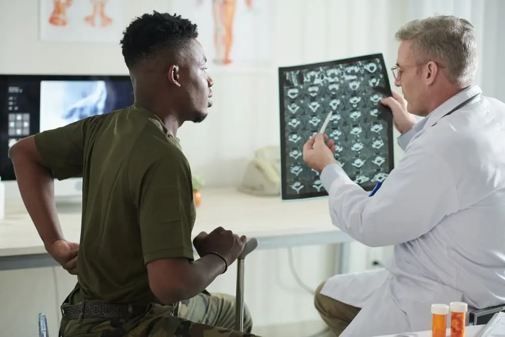
Back pain can have many possible causes, and an MRI can provide detailed images of the spine, discs, nerves, ligaments, and surrounding soft tissues when imaging is medically appropriate.
A back or lumbar spine MRI may help a medical provider evaluate conditions such as a herniated disc, spinal stenosis, nerve compression, degenerative changes, certain injuries, infection, or other abnormalities. However, not everyone with new back pain needs an MRI. Your medical provider should determine whether imaging is appropriate based on your symptoms, examination, medical history, and how your condition is progressing.
This guide explains what a back MRI can show, when one may be recommended, what happens during a lumbar MRI, and one of the most common questions patients ask us: Does your whole body go into the MRI machine for a lower back MRI?
Does Your Whole Body Go In for a Lower Back MRI?
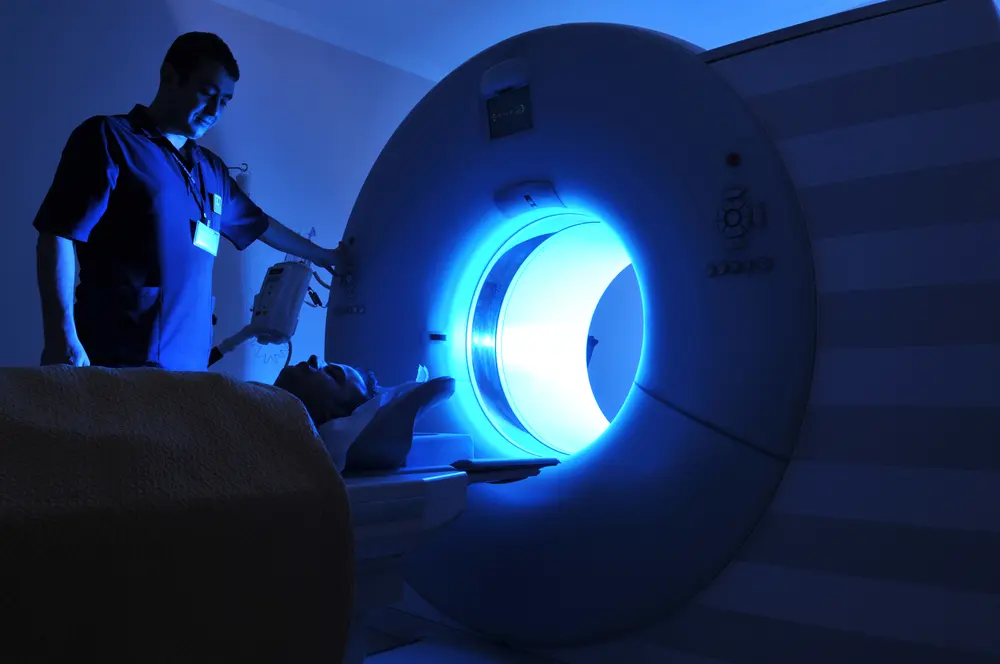
Not necessarily. During a lumbar or lower-back MRI, the lower spine must be positioned in the correct part of the scanner so clear images can be obtained. Exactly how much of your body enters the scanner depends on the MRI equipment, your height, the area being examined, and how the technologist needs to position you.
With some MRI systems, a lumbar examination can be performed feet-first and your head may remain near or outside the opening. An open MRI also provides a less enclosed design than a traditional closed-bore scanner, which many patients find more comfortable.
If you are worried about claustrophobia or how far you will enter the MRI machine, tell the imaging center before your appointment. The staff can explain the specific equipment and positioning used for your exam.
What Can an MRI Show for Back Pain?
MRI creates detailed images of the spine and surrounding tissues without using ionizing radiation. Compared with a standard X-ray, MRI provides much more information about soft tissues such as spinal discs, nerves, ligaments, the spinal cord, and surrounding structures.
A back MRI may help a medical provider evaluate abnormalities including:
Herniated or Bulging Discs
The discs between the vertebrae act as cushions in the spine. MRI can show changes in the shape and condition of these discs, including disc bulges and herniations.
A disc abnormality does not automatically mean it is responsible for someone’s pain. Imaging findings should be interpreted together with the patient’s symptoms and physical examination.
Spinal Stenosis
Spinal stenosis is narrowing of the space within the spinal canal or around spinal nerves.
MRI can help show whether narrowing is affecting the spinal canal or nearby nerve structures.
Nerve Compression
MRI provides detailed images of the structures around spinal nerves and can help identify conditions that may be compressing or irritating a nerve.
This can be particularly relevant when back pain is accompanied by symptoms such as pain traveling into a leg, numbness, tingling, or weakness.
Degenerative Changes
MRI can show age-related changes involving spinal discs, joints, and other structures.
However, degenerative changes are common and can also appear in people who do not have back pain. A medical provider should determine whether an imaging finding is actually related to a patient’s symptoms.
Injuries to the Spine and Surrounding Tissues
Depending on the circumstances, MRI can help evaluate injuries involving discs, ligaments, bone marrow, spinal cord, nerves, and surrounding soft tissues.
Infection, Tumors, and Other Less Common Conditions
MRI can also help physicians evaluate certain infections, tumors, inflammatory conditions, and other abnormalities affecting the spine or surrounding structures when those conditions are suspected.
When Might a Doctor Recommend an MRI for Back Pain?
An MRI is not usually needed for every new episode of uncomplicated lower-back pain. Many cases improve without advanced imaging.
A medical provider may be more likely to recommend spine imaging when there are specific concerns based on the patient’s symptoms, history, examination, or response to treatment.
Examples can include:
- Persistent or worsening symptoms despite appropriate care
- Progressive weakness or numbness
- Pain radiating into an arm or leg when nerve involvement is suspected
- Significant trauma
- Concern for spinal cord or nerve compression
- Previous spine surgery with new or worsening symptoms
- Concern for infection
- History or signs that raise concern for cancer
- Other clinical findings that make advanced imaging appropriate
Some Symptoms Need Prompt Medical Evaluation
Seek prompt medical attention rather than simply scheduling an imaging test yourself if back pain is accompanied by concerning symptoms such as:
- New difficulty controlling the bladder or bowels
- Progressive leg weakness or numbness
- Difficulty walking or loss of balance
- Significant recent trauma
- Fever along with back pain
- A known cancer history with new concerning back symptoms
An MRI may be part of the evaluation in some of these situations, but the appropriate next step should be determined by a healthcare professional.
What If Your MRI Is Normal but Your Back Still Hurts?
A normal or relatively unremarkable MRI does not mean that someone’s pain is imaginary or unimportant.
Back pain can occur without a single structural abnormality that clearly explains the symptoms on an MRI. Pain may involve muscles, movement, joints, inflammation, posture, or other factors that are not always represented by one obvious MRI finding.
The opposite can also occur: an MRI may show disc degeneration, bulges, or other changes in someone who has little or no pain.
For that reason, an MRI report should not be interpreted in isolation. Your healthcare provider should consider the images together with your symptoms, medical history, and physical examination.
If your MRI does not reveal a clear explanation for your symptoms, speak with the healthcare provider managing your back pain about the appropriate next steps.
Can an MRI Show a Pinched Nerve in the Back?
MRI can show the structures surrounding the spinal nerves and can help identify conditions that may compress a nerve, such as a herniated disc or narrowing around the nerve.
The MRI does not simply label every case as a “pinched nerve.” A radiologist evaluates the images for narrowing, disc abnormalities, nerve-root compression, inflammation, and other findings.
Those imaging findings should then be considered together with symptoms such as:
- Radiating leg pain
- Numbness
- Tingling
- Weakness
- Changes in reflexes
Learn more about how MRI can help evaluate a pinched nerve.
Can an MRI Show Muscles and Soft Tissues in the Back?
Yes. MRI provides detailed images of many soft tissues, including muscles, ligaments, tendons, spinal discs, nerves, and other structures surrounding the spine.
Depending on the type of injury and the MRI protocol ordered, these images can help a radiologist evaluate certain soft-tissue abnormalities.
However, not every case of muscle-related back pain produces a specific abnormality on an MRI. Imaging findings should be considered in the context of the patient’s symptoms and examination.
What Should You Expect During a Lower Back MRI?
Before the Scan
The MRI staff will review safety information with you before the exam.
Tell the staff if you have:
- A pacemaker or implanted medical device
- Metal fragments or implants
- Previous surgery
- Pregnancy or possible pregnancy
- Kidney problems if contrast may be considered
- Previous reactions to MRI contrast
- Claustrophobia or significant anxiety
You may be asked to remove jewelry, watches, removable metal objects, and certain clothing before entering the MRI area.
During the Scan
A technologist will position you on the MRI table so the part of your spine being examined is properly centered for imaging.
You will need to remain still while the images are being created.
MRI machines typically make loud tapping, knocking, or thumping sounds while images are acquired. Hearing protection is generally provided.
The technologist can monitor you during the examination and communicate with you while the scan is in progress.
How Long Does a Back MRI Take?
The exact time varies according to the type of spine MRI, equipment, imaging protocol, and whether contrast is required.
A spine MRI commonly takes approximately 30 to 60 minutes, although some examinations may be shorter or longer.
Ask the scheduling team about the expected duration of your particular scan.
Open MRI for Back Pain in Brooklyn, NY
If your medical provider has ordered an MRI for back pain, Brooklyn Open MRI provides diagnostic imaging at: 3506 Nostrand Avenue, Brooklyn, NY 11229
Our open MRI equipment provides a less enclosed imaging environment than a traditional closed-bore scanner, which may be more comfortable for patients who experience claustrophobia or anxiety.
Same-day appointments may be available depending on the examination, prescription or referral requirements, insurance authorization, and scheduling availability, or Call 718-400-6369
Frequently Asked Questions About MRI for Back Pain
Do I Need an MRI for Back Pain?
Not everyone with back pain needs an MRI. For uncomplicated new lower-back pain, imaging may not be necessary. A healthcare provider may recommend MRI when symptoms persist or worsen, neurological symptoms develop, serious underlying conditions are suspected, or imaging is needed to help guide further care.
Your healthcare provider should determine whether an MRI is appropriate for your specific symptoms.
What Does a Lower Back MRI Show?
A lumbar spine MRI creates detailed images of the lower spine and surrounding structures.
Depending on the examination, it can show:
- Vertebrae
- Spinal discs
- Spinal canal
- Nerve roots
- Ligaments
- Muscles and surrounding soft tissues
It can help identify findings such as disc herniation, spinal stenosis, nerve compression, degenerative changes, certain injuries, and other abnormalities.
Can an MRI Show a Herniated Disc?
Yes. MRI is commonly used to evaluate the spinal discs and can show whether a disc is bulging or herniated.
However, a disc abnormality on an MRI does not automatically prove that it is the source of a person’s pain. A healthcare provider should compare the imaging findings with the patient’s symptoms and examination.
INTERNAL LINK
Link “herniated disc” to:
/open-mri-accuracy-herniated-disc/
Can an MRI Show Sciatica?
MRI does not diagnose the symptom of sciatica itself, but it can help identify structural conditions that may irritate or compress a spinal nerve and contribute to sciatic symptoms.
Examples include a herniated disc or narrowing around a nerve.
Is MRI Better Than an X-Ray for Back Pain?
MRI and X-ray provide different kinds of information.
X-rays are particularly useful for evaluating bones, alignment, and certain fractures. MRI provides much more detailed images of soft tissues such as discs, nerves, ligaments, the spinal cord, and surrounding structures.
One test is not automatically “better” for every situation. Your healthcare provider should select the imaging test that best fits the clinical question.
Does Every Back MRI Use Contrast?
No, many spine MRI examinations are performed without contrast. Contrast may be appropriate in certain circumstances depending on the condition being evaluated, prior surgery, medical history, and the imaging protocol ordered.
How Long Does a Lower Back MRI Take?
A spine MRI commonly takes about 30 to 60 minutes, although examination times vary depending on the area being scanned, equipment, protocol, and whether contrast is required.
Your imaging center can provide a more specific estimate when your appointment is scheduled.
What If My MRI Looks Normal but I Still Have Back Pain?
An MRI cannot always identify a single structural cause for back pain.
Some people experience significant pain even when MRI findings are minor or normal. Others have abnormalities on MRI without experiencing symptoms.
Your healthcare provider should interpret the imaging report together with your symptoms, examination, and medical history.
Schedule a Back or Spine MRI in Brooklyn
If your healthcare provider has ordered an MRI for back pain, contact Brooklyn Open MRI to check appointment availability and confirm what documentation is required before your scan.
Brooklyn Open MRI
3506 Nostrand Avenue
Brooklyn, NY 11229
Same-day appointments may be available depending on the examination, prescription or referral requirements, insurance authorization, and scheduling availability, or Call 718-400-6369