Although Alzheimer’s disease can’t be cured, an MRI is a great tool that can show how the brain is functioning and therefore help slow the progression of the disease. Here’s what you need to know about how an Alzheimer’s disease MRI can be used to treat this illness.
What is Alzheimer’s Disease?
Alzheimer’s is a progressive neurological disease that causes the brain to shrink and neurons to die. It can lead to symptoms such as:
- Memory loss
- Wandering and getting lost
- Repeating questions
- Taking longer to complete tasks
- Poor judgment
- Changes in mood or personality
Although the exact cause of Alzheimer’s is unknown, scientists believe that it results from the buildup of thick protein deposits in the brain called beta-amyloid plaques, which causes neurons to stop working and lose connection with neighboring cells.
A person’s likelihood of developing Alzheimer’s increases with age, but individuals can begin to show signs of early-onset Alzheimer’s in their 40s and 50s. You may also be at a higher risk of developing the disease if someone in your family has it or if you suffer from heart disease or other cardiovascular issues.
How Can MRIs Diagnose Alzheimer’s Disease?
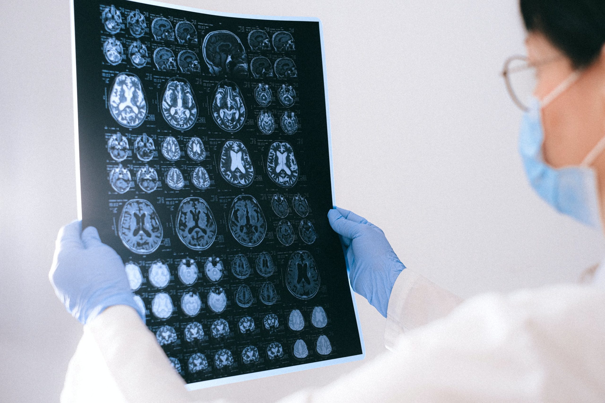
An MRI, short for magnetic resonance imaging, is a non-invasive exam that takes detailed images of the brain and surrounding tissues using powerful magnets and radio waves. Unlike CT scans or X-rays, MRIs don’t use radiation, which can be harmful to the DNA in your cells.
The 3D imaging that Alzheimer’s disease MRIs use makes it easy for doctors to locate abnormalities in the brain. One of the most commonly affected areas of the brain in Alzheimer’s patients is the hippocampus, responsible for creating and storing new memories. The parietal lobe, responsible for visual and spatial perception, is also negatively affected by Alzheimer’s.
The detailed 3D images that MRI scans take can depict how big these structures are and how many cells they contain, which can be valuable in making an accurate diagnosis. Although there is still a lot more research to be conducted, MRIs are one of the best options to diagnose Alzheimer’s disease.
What Should You Expect During an MRI?
During an MRI, you’ll be instructed to enter the scanning room and lie down on a table that slides into the imaging machine. Unlike the traditional tube-like MRI structure that can cause anxiety or claustrophobia in some patients, an open MRI in Brooklyn is a more comfortable and convenient experience because it’s available on all sides.
You’ll also be provided with headphones to tune out the sounds that often accompany an MRI scan, which can include clicking, knocking, and thumping. Don’t worry if you hear these noises throughout your exam; they’re entirely normal.
Perhaps most important of all is to stay still throughout your MRI exam. This ensures that the images will be clear and easy for your doctor to examine. If you’re feeling anxious and unable to remain still, a physician can give you an anti-anxiety medication to make you feel more comfortable.
If you’re wondering where to get an MRI in Brooklyn, look no further. At Brooklyn Open MRI, we’re committed to providing high-quality MRI diagnostic services. Don’t hesitate to reach out for help if you’re concerned about your symptoms and believe that you may have early-onset Alzheimer’s.
The importance of early diagnosis can’t be understated, as the disease is easier to treat if it’s caught in its initial stages. An MRI scan can provide your doctor with a detailed view of your brain and allow for the formulation of a comprehensive treatment plan. Schedule an appointment with Brooklyn Open MRI today.